
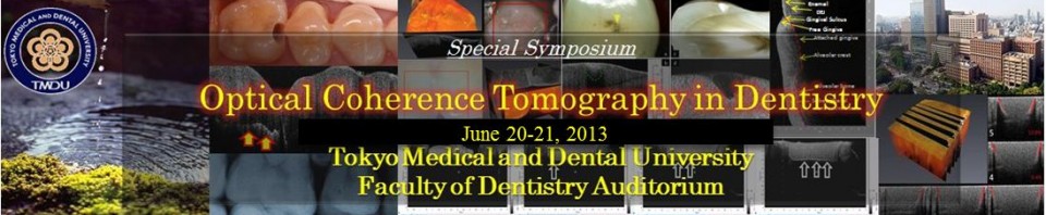
The 1st International Symposium and Mini-Exhibition
Optical Coherence Tomography in Dentistry
Date: June 20-21, 2013
Place: TMDU Faculty of Dentistry Auditorium, 4th floor
Download Abstract Book of The 1st International Symposium on Optical Coherence Tomography in Dentistry (PDF)
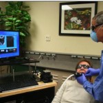
See the Symposium Photo Gallery
Organizing Committee
Prof. Junji Tagami
Dr. Yasushi Shimada
Dr. Alireza Sadr
Special thanks to TMDU Student Chapter members and Global COE Advanced International Super Students (AISS, QAISS) for their support and contribution to the program:
Dr. Turki Bakhsh
Dr. Mona Mandurah
Dr. Ikumi Wada
Dr. Baba Bista
Dr. Maria Romero
Dr. Patrycja Majkut
Dr. Kei Horie
Dr. Hiroki Tezuka
Dr. Tomoka Ueno
Dr. Juri Hayashi
Dr. Uyanga Enkhbold
Dr. Yuan Zhou
Dr. Jorge Espigares
Mini-Exhibition
The following participated in the exhibition
Kuraray Noritake Dental, Inc.
Santec Corporation
System Engineering/Axsun Technologies
Thorlabs Japan
Tokuyama Dental Corporation
TMDU Student Chapter of OSA
UTC Aerospace Systems
Symposium Chair
 Keynote Address and Welcome Message
Junji Tagami, D.D.S., Ph.D.
Professor, Cariology and Operative Dentistry
Dean, Faculty of Dentistry
Tokyo Medical and Dental University
Message
Optical coherence tomography is (OCT), is an emerging technology that can provide tomographic images of the tissue without using X-ray. This non-invasive imaging modality has a promising prospect for dentistry. It is an honor for us to have leading researchers in the field of OCT from home and abroad to discuss the applications of this technology for various purposes including non-destructive monitoring of oral structures and related topics in this symposium. We have been involved in the development of SS-OCT intra-oral probe for dental application and published several papers in the past 5 years on the great potentials of this amazing modality in our field. Researchers, students, clinicians and entrepreneurs are cordially invited to attend this symposium, which is held in the most central National Dental School in Japan, and share the excitement of the next stage of adopting OCT in dentistry.
BiographyProf. Junji Tagami received his DDS degree at the Faculty of Dentistry, Tokyo Medical and Dental University in 1980. He obtained his Ph.D. degree under the supervision of Profs. Takao Fusayama and Hiroyasu Hosoda in Dental Science in March 1984. In 1987 and 1988, he studied under Professor David Pashley, Medical College of Georgia. Currently, Dr. Tagami is Dean of the Faculty of Dentistry and Dean of Graduate School at Tokyo Medical and Dental University. His primary research interest involves studies related to the adhesion of restorative materials to tooth substance and dental materials within the broad area of adhesive dentistry, and the cariology. He has led the OCT project at Tokyo Medical and Dental University with a vision to promote this technology in Japan and globally as a tool for clinical dentistry. His research works have been published in more than 300 international and national peer-reviewed journals.
Invited Speakers
 Imaging Tooth Demineralization and Remineralization with Polarization Sensitive Optical Coherence Tomography
Daniel Fried, Ph.D.
Professor
Preventive & Restorative Dental Sciences
University of California, San Francisco
Abstract
New methods are needed for the nondestructive measurement of tooth demineralization and remineralization, to monitor the progression of incipient caries lesions and assess lesion activity. If caries lesions are detected early enough they can be arrested by chemical intervention and dietary changes without the need for surgical intervention.
Optical coherence tomography is ideally suited to monitor the changes that occur in caries lesions, since it can nondestructively image the internal structure of the lesion with an axial resolution exceeding 10-µm. Lesions can become arrested due to the preferential deposition of mineral in the outer surface zone of the lesion. The deposition creates a highly mineralized and weakly scattering surface zone that is clearly discernable in OCT images. Since this zone is near the highly reflective tooth surface, cross-polarization OCT imaging can greatly facilitate resolution of this zone. The contrast of demineralization is also increased in the cross polarization OCT images. In this presentation, the results of in vitro OCT imaging studies employing different demineralization and remineralization regimens that produce lesions with varying mineral gradients will be discussed. Automated algorithms were developed to assess the lesion depth and severity, even with highly variable mineral gradients in the lesions. The results of recent in vivo imaging studies on natural occlusal lesions, the development of early lesions, and remineralization of lesions with fluoride will also be presented. This work is supported by research grant R01-DE17869 from the NIH/NIDCR.
Dr. Daniel Fried is a professor in the Division of Biomaterials and Bioengineering in the Department of Preventive and Restorative Dental Sciences at the University of California, San Francisco School of Dentistry. He received his PhD in Physical Chemistry from Wayne State University in 1992 and his thesis work involved laser ablation and spectroscopy. Dr. Fried has been a researcher in the field of laser dentistry for the past 20 years and he has published more than 180 research publications in this field. His work has included: fundamental measurements of the optical properties of dental hard tissues from the UV to the IR, studies of the interaction of carbon dioxide lasers with dental hard tissues for laser ablation of caries and the surface modification of enamel for caries prevention, the use of lasers for the selective removal of caries and composite restorative materials, the assessment of demineralization and remineralization with polarization sensitive optical coherence tomography, and the development of near-IR imaging for caries detection. He is an editor of Journal of Biomedical Optics and SPIE conference proceedings Lasers in Dentistry.

Expanding the Domain of Optical Coherence Tomography in Dentistry
Robert Jones, D.D.S., Ph.D.
Assistant Professor
Division of Pediatric Dentistry
School of Dentistry University of Minnesota
Abstract
In this presentation, several important ‘expanded use’ dental OCT applications will be discussed. The main focus will be examining how cross polarization OCT can be a useful modality to further understand the process of secondary caries. Clinical investigation of composite resin restorations will be discussed with an emphasis on the need to sample and study biofilms associated with restorations in various stages of health and disease. Several examples from studies examining biofilm growth will be presented including a key validation study. The influence of composite formulations for OCT diagnostic imaging will also be presented. Lastly, this presentation will also discuss the challenges associated with using OCT for CAD/CAM fabrication.
Biography Dr. Jones received his DDS and PhD after completing the Dental Scientist Training Program at University of California, San Francisco. This NIH sponsored T32 training program gave Dr. Jones a graduate education in medical and optical biomedical imaging. His PhD was completed under the mentorship of Dr. Daniel Fried, whose research career has focused on lasers and optical imaging in dentistry. He completed his Pediatric Dental Residency at UCSF in 2009 and has done research in examining biological factors in Early Childhood Caries. Dr. Jones joined pediatric dentistry faculty and collaborative research environment at the University of Minnesota in 2009. Dr. Jones is a 2010 recipient of the Faculty Development Award from the 3M Foundation. He is also a Co-PI on a funded NIH grant (Sept 2010) examining the interaction of oral biofilms and dental resin composites. He is a pioneer of dental OCT with numerous key publications on the basics and applications of the technology. His current research interests include early caries detection using near infrared based imaging, e.g. OCT, and assessing risk factors in early childhood caries.
 Development of a New Oral Diagnostic Tool using Optical Coherence Tomography
Yasunori Sumi, D.D.S., Ph.D.
Professor and Director
Department for Advanced Dental Research
Center of Advanced Medicine for Dental and Oral Diseases
National Center for Geriatrics and Gerontology (Japan)
Abstract
Optical coherence tomography (OCT) is a new biomedical imaging modality which can generate high-resolution, cross-sectional images of microstructures in biological systems. One of the most attractive features of OCT is that it uses safe near-infrared light instead of hazardous ionizing radiation. Furthermore, resolution on the order of 10 micrometers can be obtained. The optical accessibility of clinically relevant structures in the oral cavity makes it a particularly attractive location for the application of OCT imaging techniques. Our National Center for Geriatrics and Gerontology has developed a new swept-source optical coherence tomography (SS-OCT) system by industry and public-sector joint research. This new SS-OCT system was applied for cross-sectional imaging of dental caries, resin based composite restorations, periodontal disease, oral cancer and finished dentures in this review. It is concluded that our new SS-OCT system is a promising new and useful alternative imaging technique which can safely provide much more definitive information on oral structures at far higher resolution than possible by conventional clinical imaging methods. Our National Center for Geriatrics and Gerontology has this new dental SS-OCT system of Japanese origin into the world’s first production.
Biography Dr. Sumi received his DDS from TMDU in 1981 and completed post-graduate course in oral surgery at Nagoya University in 1985. His primary research interest has focused on oral care for the elderly. He is the pioneer of development of dental OCT in Japan, and has worked with a number of co-investigators in the OCT project funded by Research Grant for Longevity Sciences from Ministry of Health, Labor and Welfare. Under his direction, dental SS-OCT system has been developed at National Center for Geriatrics and Gerontology of Japan. He holds several international patents related to dental OCT imaging. Dr. Sumi is also the editor-in-chief of Journal of the Japanese Society of Gerodontology and serves on the editorial board of Geriatrics & Gerontology International.
Distinguished Speakers

NIR Imaging of Dental Decay
Cynthia Lee Darling
Associate Professor
Preventive & Restorative Dental Sciences
University of California, San Francisco
Abstract
Near-Infrared (NIR) imaging is a new technology that is currently being investigated for the detection and assessment of dental caries. Recent In vivo and in vitro imaging studies have shown that high contrast images of tooth demineralization can be acquired in the NIR due to the high transparency of dental enamel and that the maximum contrast between sound and demineralized enamel lies in the 1300-1600-nm region. Therefore, this wavelength range is well suited for transillumination and reflectance imaging of dental caries. Images of the lesion can be acquired from the facial, lingual, and occlusal surfaces and from multiple angles for optimum viewing. Many of the chromophores responsible for stains do not absorb light in the NIR allowing for easier discrimination of carious lesions in occlusal surfaces. This method also has great potential for the examination of defects in tooth structure, and internal cracks in the enamel are visible due to the high transparency of the enamel in the NIR. Our recent clinical study showed that NIR imaging has great potential as a screening tool for the detection of occlusal and approximal lesions without the use of ionizing radiation (x-rays). Support: NIH Grant R01-DE14698.
Biography
Cynthia Lee Darling is an Associate Professor in the Division of Biomaterials and Bioengineering in the Department of Preventive and Restorative Dental Sciences at the University of California, San Francisco School of Dentistry. She received her PhD in Physical Chemistry from Wayne State University in 1993 in the area of theoretical quantum and classical mechanics. Dr. Darling has been a researcher in the field of biomedical photonics for the past eleven years and her contributions made to this field include development of a fully automated Mueller polarimetric imaging system to completely describe the interaction of polarized light with dental hard tissues. Dr. Darling and her research group have also investigated the optical properties of developmental defects, employed NIR imaging to monitor laser ablation through dental enamel in real-time to directly visualize peripheral thermal and mechanical damage, and explored the image contrast of dental caries at other NIR wavelengths besides 1300-nm.
 Early Dental Caries Assessment with Optical Coherence Tomography and Polarized Raman Spectroscopy
Lin-P’ing Choo-Smith, Ph.D.
Research Officer
National Research Council Canada
Abstract
In recent years, we have been exploring the use of optical coherence tomography (OCT) and polarized Raman spectroscopy (PRS) as potential clinical tools for in vivo early dental caries assessment. The underlying basis is that OCT and PRS furnish complementary morphological depth imaging and biochemical specificity, respectively, to provide clinicians with an improved detection tool over conventional clinical methods. This presentation will describe our research results ranging from early laboratory bench studies on extract human tooth samples towards the development of portable and specialized OCT and PRS prototype systems and fibre-optic intra-oral probes for in vivo assessment with patient volunteers. The strengths, limitations and new insights from combining OCT and PRS for dental caries assessment will also be discussed.
Biography Dr. Lin-P’ing Choo-Smith is a Research Officer in the Medical Devices Portfolio at the National Research Council Canada (NRC; Winnipeg, MB). She has a B.Sc. from the University of Toronto (1992), Ph.D. from the University of Manitoba (1996) and post-doctoral training at Case Western Reserve University (Cleveland, OH, USA; 1996-1997) and Erasmus University Rotterdam (Rotterdam, The Netherlands; 1997-2001). Her research interests include biomedical applications of Raman spectroscopy and applied photonics especially dental caries assessment. Since joining the NRC in 2001, she has been involved with developing the combination of optical coherence tomography and Raman spectroscopy for early dental caries detection and monitoring. This research involves dental clinical collaborators from the Faculty of Dentistry at the University of Manitoba and Dalhousie University. Dr. Choo-Smith is the principal investigator of a research team with funding over the years through NRC and grants from the Canadian Institutes of Health Research and US National Institutes of Health.
TMDU Speakers
SS-OCT for the detection of dental caries and tooth crack
Yasushi Shimada, D.D.S., Ph.D.
Cariology and Operative Dentistry
Department of Oral Health Sciences
Tokyo Medical and Dental University
Abstract
Caries diagnosis: The diagnosis of dental caries of posterior teeth is a challenge due to the restricted access for examination. Despite the dramatic decline of caries incidence, the disease is far from eradicated, particularly in children and young adults. The occlusal fissures of the first permanent molars are generally the first sites in the permanent dentition to develop caries. Although it is well accepted that bitewing radiography provides additional benefits in the detection of occlusal caries and non-cavitated proximal lesions, evidence for their value in epidemiological studies is still controversial. Dental radiographs seem to exhibit low sensitivity, but rather high specificity, while the true extent of caries lesions seems to be underestimated when relying solely on radiographs. From our in vivo and in vitro studies, Swept Source OCT (SS-OCT) showed higher sensitivity and Az values of ROC analysis than the conventional caries detection method, especially for the detection of cavitated enamel lesions and dentin caries.
Tooth crack diagnosis: Cracked teeth have been a diagnostic challenge for more than half century because of the difficulty in locating crack lines of incomplete tooth fracture. Most clinicians may not find the presence of crack until they encounter complicated and diverse symptoms associated with chewing and cold. Longitudinal tooth fractures have been categorized into 5 major classes: craze line, fractured cusp, cracked tooth, split tooth, and vertical root fracture (Cracking the cracked tooth code, American Association of Endodontics). Since a crack has an unpredictable prognosis including extraction, accurate diagnosis regarding the size and localization of the crack is required to determine the most appropriate treatment technique.SS-OCT was employed for the detection of naturally formed cracks and was capable of providing clear imaging of enamel cracks including information on penetration depth. Even when cracks extended beyond DEJ, OCT was capable of imaging the whole line, determining crack penetration depth.
Biography Dr. Shimada is a senior faculty member of Cariology and Operative Dentistry. He studied dentistry and took his Ph.D. at the same institution. His extended research activities have involved characterization of dental adhesives, introducing new methodologies such as the wire-loop micro-shear bond strength test. Dr. Shimada has the experience of working at the National Institute of Standards and Technology (NIST), and has also been involved in research on pulp biological response. He is currently involved in the OCT research project with a focus on the clinical aspect, and development of an intra-oral imaging probe for chair-side imaging.
 Time-resolved and Quantitative Analysis of Dental Structures by Optical Coherence Tomography
Alireza Sadr, D.D.S., Ph.D.
Cariology and Operative Dentistry
Department of Oral Health Sciences
Tokyo Medical and Dental University
Abstract
Swept source optical coherence tomography without polarization sensitivity has shown a unique capability for real-time imaging of dental structures, especially for time-resolved determination of defect formation.
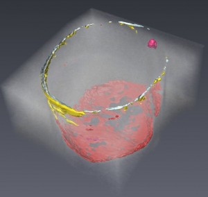
In the first part of this talk, we present our methodology for non-destructive evaluation of marginal and internal microgaps in three-dimensions as well as real-time imaging of gap formation during composite placement and polymerization. This methodology is based on the optical contrast between the media filling the defects and the composite resin or dental hard tissue which results in a detectable reflectivity signal peak. The development and evolution of this methodology has now enabled 3D visualization without the need for staining or dye penetration. Our new approach and its findings alone can establish OCT as an invaluable tool for dental materials research; however, there are still limitations that should not be over looked such as imaging depth and variations among composites. In the latter part of this talk, we int
roduce our latest clinical results on quantitative monitoring of enamel lesion remineralization based on OCT signal analysis. In the series of case presented, enamel white-spot lesions were detected in patients and monitored over time while the patients were exposed to increased levels of salivary Ca and F through daily consumption of a chewing gum (POs-Ca F). Successful healing of some lesions are evident from OCT images obtained as soon as one month after the treatment started; nevertheless, the degree of success was varied among different cases.
Dr. Sadr is a Junior Associate Professor at Cariology and Operative Dentistry, Department of Oral Health Sciences. He joined TMDU as a graduate student in 2003, received his Ph.D. as a Japanese government scholar under supervision of Prof. Tagami in 2008 and became a faculty member and principal investigator at the Global COE program in 2009. His field of research involves characterization of dental hard tissue and biomaterials. He is also enthusiastically working on the OCT development project with Ph.D. candidates and colleagues, investigating alternative methodologies for OCT image and signal analyses, monitoring dental lesions and defects. He has authored and coauthored over 80 full-length research publications with his colleagues at TMDU, and received funding for several research projects from the Japanese Society of Promotion of Science (JSPS) and Japanese Ministry of Education (MEXT). His research has brought him several domestic and international scientific awards including three consecutive outstanding research prizes in Adhesive Dentistry in 2011, 2012 and 2013.
Schedule of the Events
Day 1- June 20, 2013 (Thursday)
| Time |
Subject |
Speaker/Remarks |
| 09:00-10:00 |
Poster and Exhibition Setup, Registration Open
|
|
| 10:00-10:45 |
Keynote Address and Welcome Message |
Junji Tagami |
| 10:45-11:30 |
Imaging Tooth Demineralization and Remineralization with Polarization Sensitive OCT |
Daniel Fried |
| 11:30-12:15 |
Expanding the Domain of Optical Coherence Tomography in Dentistry
|
Robert Jones |
| 12:15-13:00 |
Exhibition and Poster Viewing (Lunch Break ) |
|
| 13:00-13:45 |
Development of a New Oral Diagnostic Tool using OCT |
Yasunori Sumi |
| 13:45-14:15 |
SS-OCT for Caries and Crack Detection |
Yasushi Shimada |
| 14:15-14:45 |
Time-resolved and Quantitative Analysis of Dental Structures by OCT |
Alireza Sadr |
| 14:45-15:30 |
Exhibition and Poster Viewing (Coffee Break) |
|
| 15:30-15:40 |
Solutions for SS-OCT using Santec Technology |
A. Morosawa (Santec Corp.) |
| 15:40-15:50 |
High line rate 2048 pixel InGaAs camera for SD-OCT @1.05/1.31 μm |
D. Malchow (UTC Aerospace Systems) |
| 15:50-16:00 |
Science of Supra-Nano Filled Composites |
T. Suzuki (Tokuyama Dental Corp.) |
| 16:00-16:10 |
Swept Lasers Designed for Biomedical Imaging Applications |
W. Ahern (Systems Engineering and Axsun Technologies) |
| 16:10-16:30 |
Group Discussions and Closing of the First Day |
|
|
18:00-20:00
|
Dinner Reception |
Invitation Only |
Day 2- June 21, 2013 (Friday)
|
| Time |
Subject |
Speakers |
| 9:00-10:00 |
Registration Open |
|
| 10:00-10:30 |
Near Infrared Imaging of Dental Decay |
Cynthia Darling |
| 10:30-11:00 |
Assessment of Early Enamel Erosion with OCT |
Hooi Pin Chew |
| 11:00-11:15 |
Coffee Break (Poster Viewing) |
|
| 11:15-12:00 |
Early Dental Caries Assessment with Optical Coherence Tomography and Polarized Raman Spectroscopy |
Lin-P’ing Choo-Smith |
| 12:00-12:30 |
Laboratorial Method Proposal to Obtain Caries-affected Dentin Observed by OCT |
Adriana Bona Matos |
| 12:30-13:15 |
Lunch Break |
|
| 13:15-13:35 |
A Brief Review on Quantitative Diagnosis Technologies for Early Carious Lesions |
Syozi Nakashima |
| 13:35-13:50 |
Marginal Adaptation of Self-etch Adhesives by 3D Optical Coherence Tomography |
Patricia Makishi |
| 13:50-14:05 |
Estimation of the Enamel and Dentin Mineral Content from the Refractive Index |
Ilnaz Hariri |
| 14:05-14:20 |
Relationship between OCT image, Microscopic Gap and Bond Strength of Composites |
Turki Bakhsh |
| 14:20-14:35 |
Assessment of Tooth Fracture using SS-OCT |
Yukie Nakajima |
| 14:35-14:45 |
Closing |
|
|
Poster Presentations
|
| Poster No. |
Title |
Presenter |
| 101 |
Optical and Nano-indentation Mechanical Properties Evaluation of Enamel Coated by Resin-thin-film |
Ehab Alsayed |
|
102
|
Non-destructive Assessment of Current One-step Self-etch Dental Adhesives using Optical Coherence Tomography |
Baba Bista |
| 103 |
Comparison of Optical Coherence Tomography versus micro-CT for visual assessment of early enamel lesions |
Jorge Espigares |
| 104 |
Assessment of Non-carious Cervical Lesions using Optical Coherence Tomography |
Ikumi Wada |
| 105 |
Characterization of Transparent Dentin in Attrited Teeth using Optical Coherence Tomography and Nanoindentation |
Mona Mandurah |
| 106 |
Sealing performance of resin cements monitored using optical coherence tomography |
Alaa Turkistani |
| 107 |
Assessment of remaining dentin thickness during caries excavation by SS-OCT |
Patrycja Majkut |
| 108 |
Swept Lasers Designed for Biomedical Imaging Applications |
Bill Ahern |

 Early Dental Caries Assessment with Optical Coherence Tomography and Polarized Raman Spectroscopy
 In the first part of this talk, we present our methodology for non-destructive evaluation of marginal and internal microgaps in three-dimensions as well as real-time imaging of gap formation during composite placement and polymerization. This methodology is based on the optical contrast between the media filling the defects and the composite resin or dental hard tissue which results in a detectable reflectivity signal peak. The development and evolution of this methodology has now enabled 3D visualization without the need for staining or dye penetration. Our new approach and its findings alone can establish OCT as an invaluable tool for dental materials research; however, there are still limitations that should not be over looked such as imaging depth and variations among composites. In the latter part of this talk, we introduce our latest clinical results on quantitative monitoring of enamel lesion remineralization based on OCT signal analysis. In the series of case presented, enamel white-spot lesions were detected in patients and monitored over time while the patients were exposed to increased levels of salivary Ca and F through daily consumption of a chewing gum (POs-Ca F). Successful healing of some lesions are evident from OCT images obtained as soon as one month after the treatment started; nevertheless, the degree of success was varied among different cases.